Multiple Frequency Tympanometry
Tympanometry Guides
Overview
Tympanometry performed with a probe tone frequency of 226 Hz provides useful test mode clinical information regarding disorders of the tympanic membrane and the Eustachian tube. The middle-ear system is mainly stiffness-controlled when tympanometry is performed with low frequency probe tones. Ossicular abnormalities affecting the mass-controlled components cause changes in the transmission characteristics of the tympano-ossicular system which are more easily identified with probe tone frequencies that approximate or exceed the resonance frequency of the ear.
It has been demonstrated that as the probe frequency increases, Susceptance (or the reactance component of admittance) progresses from positive values toward zero. The middle-ear system becomes less stiffness-dominated and more mass-controlled. Resonance of the system is reached when stiffness and mass components are equal in magnitude.

Tympanometric shapes progress through a series of patterns and become more complex as the probe tone frequency increases. Three patterns can be identified as follows:
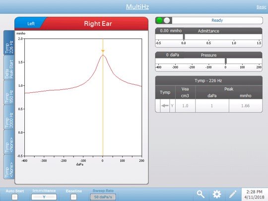
1) Low Frequency Probe Tones (Below resonance frequency): An inverted “V” shape with a single peak appearing at peak pressure.
2) Mid-Range Frequencies (The region of resonance frequencies): Multi-peaks (notching) appear and gradually evolve into an inverted W shape.
NOTE: Studies with normal subjects indicate a resonance frequency range of 600 Hz to 1340 Hz with a mean of approximately 1000 Hz (Colletti, 1977), or 800 Hz to 1200 Hz (Shanks, 1984).
3) High Frequency Probe Tones (Above resonance frequency): A “V” shape with a single peak appearing at peak pressure - an inversion of the low frequency tympanogram.
Identifying the frequency region at which these characteristic tympanometric patterns appear offers a sensitive diagnostic tool for differentiating pathological conditions of the middle-ear. The frequency values at which notching of the tympanogram appears is shifted towards higher frequencies for ossicular fixations. There may be some overlap with normal ranges. Multi-frequency investigations reveal a shifting towards lower frequencies for post-stapedectomized ears and ossicular discontinuities. Little or no overlap exists between multiple frequency patterns for ossicular fixations and interruptions.